Introduction
Background
Candidosis describes a group of yeastlike fungal infections involving the skin and mucous membranes. Infection is caused by Candida species, typically, Candida albicans. C albicans is ubiquitous and is found mainly on oral or genital mucosae; it may also be transmissible between consorts.1
By tradition, the most commonly used classification of oral candidosis divides the infection into 4 types including (1) acute pseudomembranous candidosis (thrush), (2) acute atrophic (erythematous) candidosis, (3) chronic hyperplastic candidosis, and (4) chronic atrophic (erythematous) candidosis.
Chronic hyperplastic candidosis was further subdivided into 4 groups based on localization patterns and endocrine involvement including (1) chronic oral candidosis (candidal leukoplakia), (2) endocrine candidosis syndrome, (3) chronic localized mucocutaneous candidosis, and (4) chronic diffuse candidosis.
Thrush (acute pseudomembranous candidiasis) is the term used for the multiple white-fleck appearance of acute candidiasis, which purportedly resembles the appearance of the bird with the same name.
Erythematous candidosis is the term used for the red lesions of candidiasis.
Pathophysiology
C albicans is the predominant causal organism of most candidosis. Other species, including Candida krusei, have appeared in persons who are severely immunocompromised. Candida glabrata is an emerging cause of oropharyngeal candidosis in patients receiving radiation for head and neck cancer.2 In patients with HIV infection, new species, such as Candida dubliniensis and Candida inconspicua, have been recognized.
C albicans is a harmless commensal organism inhabiting the mouths of almost 50% of the population (carriers); persister cells are clinically relevant, and antimicrobial therapy selects for high-persister strains in vivo.3
Under certain circumstances, C albicans can become an opportunistic pathogen. Such a suitable circumstance for it to become an opportunist may be a disturbance in the oral flora or a decrease in immune defenses.
Acute pseudomembranous candidosis (thrush)
Thrush may be observed in healthy neonates or in persons in whom antibiotics, corticosteroids, or xerostomia disturb the oral microflora. Oropharyngeal thrush occasionally complicates the use of corticosteroid inhalers. Immune defects, especially HIV infection, immunosuppressive treatment, leukemias, lymphomas, cancer, and diabetes, may predispose patients to candidal infection.
Erythematous candidosis
Erythematous candidosis may cause a sore red mouth, especially of the tongue, in patients taking broad-spectrum antimicrobials. It also may be a feature of HIV disease. Median rhomboid glossitis is a red patch occurring in the middle of the dorsum in the posterior area of the anterior two thirds of the tongue and especially is observed in smokers and in those with HIV disease.
Chronic mucocutaneous candidosis
Chronic mucocutaneous candidosis (CMC) describes a group of rare syndromes, which sometimes include a definable immune defect, in which persistent mucocutaneous candidosis responds poorly to topical treatment. Generally, the more severe the candidosis, the greater the likelihood that immunologic defects (particularly of cell-mediated immunity) can be identified. Recent studies suggest a defect in cytokine (interleukin 2 and interferon-g) production in response to candidal and some bacterial antigens, with reduced TH1 lymphocyte function and enhanced TH2 activity (and increased interleukin 6), and reduced serum levels of immunoglobulin G2 and immunoglobulin G4.
Frequency
United States
Candidosis is common in groups at risk, such as patients who are immunocompromised. Frequency of infection is rising, primarily because of HIV infection and both the increase in candidal species other than C albicans and the resistance to antifungals.
International
Candidosis is common in groups at risk, such as patients who are immunocompromised.
Mortality/Morbidity
Candidosis may predispose individuals to esophageal spread.
Sex
Candidosis is reported equally in males and females worldwide, except in areas where males with HIV infection outnumber females.
Age
Candidosis predominantly occurs in middle-aged or older persons; however, in those with HIV infection, candidal infection primarily occurs in the third and fourth decades.
Clinical
History
Thrush
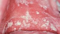
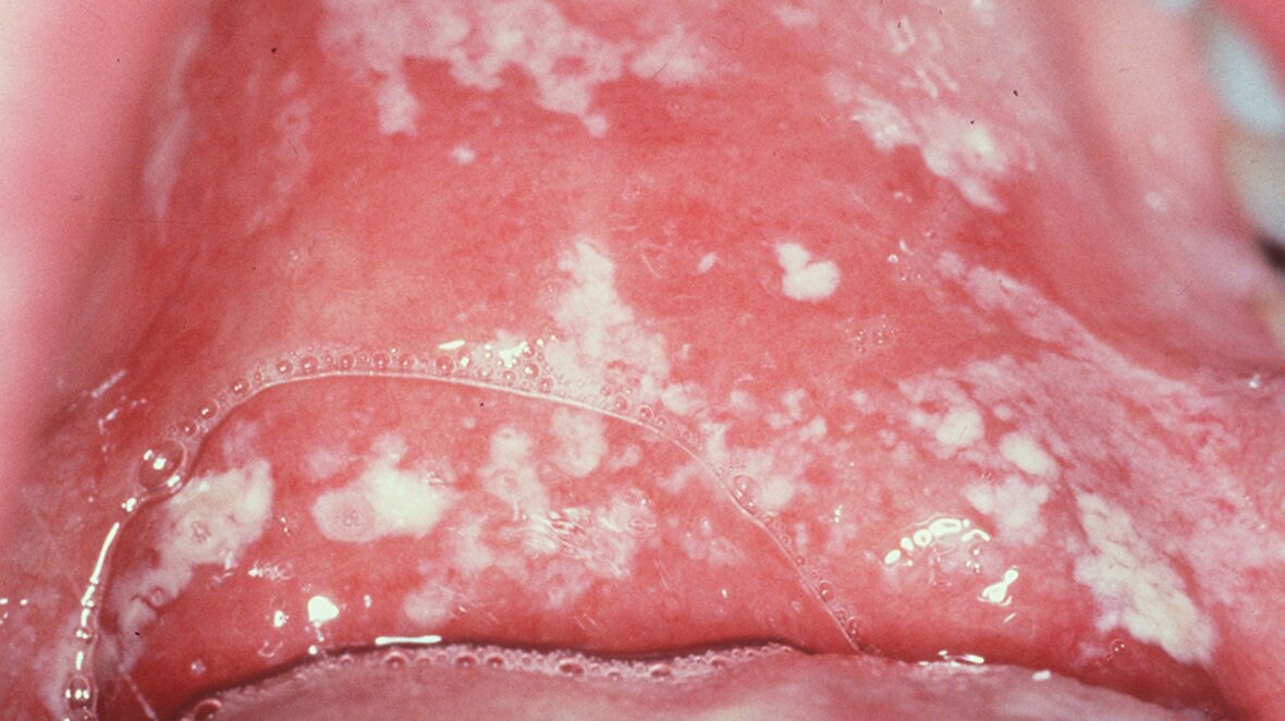
White patches on the surface of the oral mucosa, tongue, or other parts of the body characterize thrush. Lesions develop into confluent plaques that resemble milk curds and can be wiped off to reveal a raw erythematous and sometimes bleeding base. Note the image below.
Pseudomembranous candidosis.
Erythematous candidosis
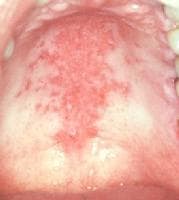
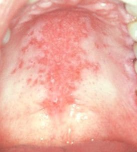
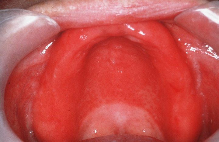
Erythematous areas found generally on the dorsum of the tongue, palate, or buccal mucosa are characteristic. Lesions on the dorsum of the tongue present as depapillated areas. Red areas often are seen on the palate of individuals with HIV infection. An associated angular stomatitis may be present. Note the image below.
Erythematous candidosis in HIV/AIDS.
Chronic hyperplastic candidosis (candidal leukoplakia)4
A chronic, discrete, raised lesion that may vary from a small, palpable, translucent, or whitish area to a large, dense, opaque plaque that is hard and rough to the touch (plaquelike lesion) is observed. Homogeneous or speckled areas, which do not rub off (nodular lesions), can be seen. Speckled leukoplakia accounts for 3-50% of candidal leukoplakias. Candidal leukoplakias usually occur on the inside surface of one or both cheeks; they occur less commonly on the tongue.
Chronic multifocal oral candidosis
In a minority of individuals, chronic candidal infection may be seen in multiple oral sites with various combinations including (1) angular stomatitis, which is unilateral or bilateral and is encountered mostly in denture wearers; (2) retrocommissural leukoplakia, which is the most constant component of the tetrad; (3) median rhomboid glossitis; and (4) palatal lesions.
Additional criteria include (1) lesions of more than 1-month duration; (2) absence of predisposing medical conditions; (3) exclusion of individuals undergoing radiotherapy or administration of the following types of drugs: anti-inflammatory, immunosuppressive, cytotoxic, or psychotropic agents or antibiotics.
This type is most common in male tobacco smokers in their fifth or sixth decade. Antifungal therapy clears the infection and produces clinical improvement; however, recurrence is common, unless smoking can be reduced.
Denture-related stomatitis (denture-induced stomatitis, denture sore mouth, chronic atrophic candidosis)5
Chronic erythema and edema of the mucosa that contacts the fitting surface of the denture are characteristic. The mucosa below the lower denture rarely is involved. Occasional slight soreness is experienced; however, the patient typically is asymptomatic. The typical presenting complaint is angular stomatitis. Note the image below.
Denture-related stomatitis; a common form of oral candidiasis. From Scully C, Flint SF, Bagan JV, Porter SR, Moos K. Atlas of Oral and Maxillofacial Diseases. 2010. Informa, London.

Denture-related stomatitis is classified into 3 clinical types as follows:
- Localized simple inflammation or a pinpoint hyperemia
- Erythematous or generalized simple type presenting as more diffuse erythema involving a part of, or the entire, denture-covered mucosa
- Granular type (inflammatory papillary hyperplasia) commonly involving the central part of the hard palate and the alveolar ridge
Angular stomatitis (perlèche, angular cheilitis)
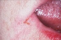
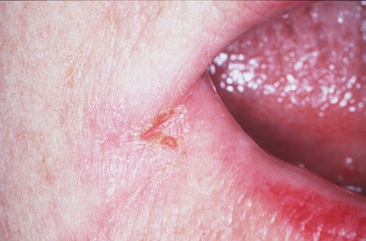
Lesions affect the angles of the mouth; soreness, erythema, and fissuring are characteristic; diagnosis commonly is associated with denture-related stomatitis. Both yeasts (candidal) and bacteria (especially Staphylococcus aureus) may be involved. Note the image below.
Angular stomatitis; a common form of oral candidiasis, typically seen in patients with denture-related stomatitis, especially those in whom the denture needs adjustment. In others, it may be a sign of diabetes, nutritional deficiency, or immune defect.
Angular stomatitis commonly is an isolated initial sign of anemia or vitamin deficiency, such as vitamin B-12, and resolves when the underlying disease has been treated. Iron deficiency anemia and other vitamin deficiencies have been cited as other predisposing factors.
In uncommon conditions, such as orofacial granulomatosis, as many as 20% of individuals have angular stomatitis, although candidal species often are not isolated. Angular stomatitis also may be seen in individuals with HIV infection.
Median rhomboid glossitis (glossal central papillary atrophy)
Papillary atrophy, which is symmetric and elliptic or rhomboidal in shape, is located centrally at the midline of the tongue, anterior to the circumvallate papillae. Occasionally, median rhomboid glossitis presents with a hyperplastic exophytic or lobulated appearance.
Histopathologically, candidal hyphae infiltrate the superficial layers of the parakeratotic epithelium, and a polymorphonuclear leukocyte infiltrate occupies the epithelium, with elongated hyperplastic rete ridges and a lymphocyte infiltration in the corium. However, the condition frequently shows a mixed bacterial-fungal microflora, as has been documented.
Other
Exfoliative cheilitis may occasionally be associated with Candida species, especially in individuals with HIV infection.
Physical
The diagnosis usually is made based on physical examination. Gram stain of a smear (hyphae) or oral rinse may aid in the diagnosis. Differentiate pseudomembranous candidosis from lichen planus. Hairy leukoplakia, leukoplakia, or Fordyce spots occasionally cause confusion. Differentiate erythematous candidosis from other inflammatory stomatitides, lichen planus, and erythroplakia.
Causes
Members of the genus Candida are the causal organisms of candidosis. Secretion of antimicrobial proteins and peptides is decreased in saliva of patients with oral candidosis.6 The following factors affect candidal carriage and infection:
- Carriage is more frequent in females than in males; carriage is frequent during the summer months.
- Increased carriage rates are seen in immunocompromised states (eg, HIV infection), blood group O, and nonsecreting of blood group antigens in the saliva possibly mediated by an effect on C albicans adhesion to epithelia.
- Carriage of yeast is higher in acidic saliva.
- Xerostomia increases the carriage of C albicans.
- Use of psychotropic drugs that cause xerostomia increases carriage of candidal organisms and S aureus.
- Candidal counts increase during sleep but are reduced by eating a meal and by brushing the teeth. Counts usually are highest first thing in the morning; the organism frequently cannot be isolated when counts are low, except in the early morning. Early morning saliva sample is the most dependable for making a comparison of the candidal population in individuals.
- Denture-wearing habits affect candidal growth. C albicans counts consistently are low in early morning saliva specimens from edentulous patients not wearing dentures. This is attributed to sleeping without dentures and the consequent alteration in the oral environment. When dentures are worn at night, the early morning saliva candidal count is high; when dentures are not worn at night, the early morning count is the lowest. Increased candidal count following reinsertion of the dentures suggests that plaque on the dentures harbors C albicans. Increase in both the frequency of carriage and the density of candidal colonization in denture wearers compared with dentate individuals suggests that prostheses encourage the presence and growth of candidal species.
- Smoking affects candidal infection. Some studies have reported that smoking significantly increased carriage from 30-70%. Smoking increased the risk in persons with HIV infection. Smoking commonly underlies multifocal candidosis and median rhomboid glossitis.
- Tetracycline therapy affects candidal growth. Candida species can be isolated from the oral cavity with greater prevalence and in greater numbers during tetracycline therapy.
- Disruption of the ecologic balance disruption can affect growth patterns. Under normal circumstances, it appears unlikely that candidal organisms establish in the mouths of noncarriers; however, if the ecologic balance is altered (by bacterial suppression, alteration of salivary flow, or immunologic deficit), candidal infection may occur.
- Similarities between carriers and noncarriers of C albicans with respect to age, caries experience, periodontal status, and intraoral temperature indicate that these factors do not influence candidal carriage significantly.
Factors predisposing individuals to oral candidal infections are as follows:
- Broad-spectrum antimicrobial therapy may predispose individuals to stomatitis or glossitis caused by C albicans.
- Topical, systemic, and aerosolized corticosteroid use may result in oral yeast infection.
- Smoking predisposes individuals to chronic atrophic candidosis and other forms of candidosis.
- Drugs with xerostomic adverse effects (eg, psychopharmaceuticals) are associated with oral candidosis. Xerostomia (as in Sjögren syndrome and after radiotherapy) predisposes individuals to candidosis.
- Immunologic disorders may play a role. Candidosis is common in patients with HIV infection and other secondary immunodeficiencies, including blood dyscrasias, diabetes, and malignant disease.
- CMC can be a feature of primary immune defects such as severe combined immune deficiency syndrome.
- Diabetes may predispose individuals to candidosis.

I was diagnose with genital warts since 2012 i have be taking lot treatment and all i got is outbreak. in 2015 I gave up the treatment because I can't continues wasting time and money on treatment at the end it will not cure me. about 6 weeks ago i did natural research online I had So many people talking good about natural remedy, after the research i was recommended to Dr onokun, And I wrote to him through his email and told him my problem after some conversations with him he gave me natural treatment after 1 week Dr onokun treated me i got cured permanently. and i went to see my doc he confirmed that the diseases has gone out from my body. every patients should know there is 100% natural hpv cure. contact Dr onokun his email address: dronokunherbalcure@gmail.com
Trả lờiXóa