Introduction
Background
The World Health Organization (WHO) first defined oral leukoplakia as a white patch or plaque that could not be characterized clinically or pathologically as any other disease; therefore, lichen planus, candidiasis, and white sponge nevus were excluded. At a 1983 international seminar, the following definition was proposed:
Leukoplakia is a whitish patch or plaque that cannot be characterized clinically or pathologically as any other disease, and is not associated with any physical or chemical causative agent, except the use of tobacco.
A more recent WHO workshop1 recommended abandoning the distinction between the terms "potentially malignant lesions" and "potentially malignant conditions" and to use the term "potentially malignant disorders" instead. Leukoplakia and erythroplakia are the most common potentially malignant disorders. These diagnoses are still defined by exclusion of other known white or red lesions.
Oral white lesions include leukoplakias (as defined above), keratoses, leukoplakias of clear infective origin (candidal, syphilitic, hairy leukoplakia associated with Epstein-Barr virus), candidosis, lichen planus, oral submucous fibrosis, lupus erythematosus, congenital lesions (eg, white sponge nevus, dyskeratosis congenita, pachyonychia congenita), and frank carcinomas.
Pathophysiology
No etiologic factor can be identified for most persistent oral white plaques (ie, idiopathic leukoplakia). The histopathologic features are highly variable, ranging from hyperkeratosis and hyperplasia to atrophy and severe dysplasia.
Patients with idiopathic leukoplakia have the highest risk of developing cancer. In studies of these patients, 4-17% had malignant transformation of the lesions in less than 20 years. The risk of developing malignancies at lesion sites is 5 times greater in patients with leukoplakia than in patients without leukoplakia.
Dysplastic lesions do not have any specific clinical appearance; however, where erythroplakia is present, dysplasia is likely.
Dysplasia is evident in 17-25% of biopsy samples of leukoplakias. Erythroleukoplakias, verrucous leukoplakias, and nodular leukoplakias show an increasing frequency of dysplastic histologic changes or aneuploidy.
Leukoplakias that are speckled, or erythroleukoplakic, are usually dysplastic or frank carcinomas. Nodular or verrucous lesions are also sinister, but homogenous leukoplakias are far less likely to be potentially malignant.
Most idiopathic leukoplakias are homogenous leukoplakias and show little evidence of dysplastic histologic changes or aneuploidy. However, studies have revealed carcinoma or severe dysplasia in the excision specimens of approximately 5% of leukoplakias excised when the diagnostic biopsy specimens had revealed no dysplasia.
Carcinoma in situ is a controversial term used for severe dysplasia in which the abnormalities extend throughout the thickness of the epithelium. All the cellular abnormalities characteristic of malignancy may be present; only invasion of the underlying connective tissue is absent. Top-to-bottom epithelial dysplasia, like other dysplastic lesions, has no characteristic clinical appearance, although erythroplasia often proves to be carcinoma in situ or early invasive carcinoma.2
Frequency
United States
Oral leukoplakia is uncommon, possibly occurring in less than 1% of adults.
International
The rates are the same as those in the United States.
Mortality/Morbidity
Some leukoplakias culminate in oral squamous cell carcinoma (OSCC).3,4
Race
An increased prevalence is observed in communities and races with high tobacco use, such as Southeast Asia.
Sex
Males have the highest incidence of leukoplakias.
Age
Leukoplakias are usually seen in adults older than 40 years.
Clinical
History
Leukoplakias are usually asymptomatic and are initially noticed by a dentist during a routine examination.
Physical
Leukoplakias are white lesions that cannot be removed with a gauze swab.
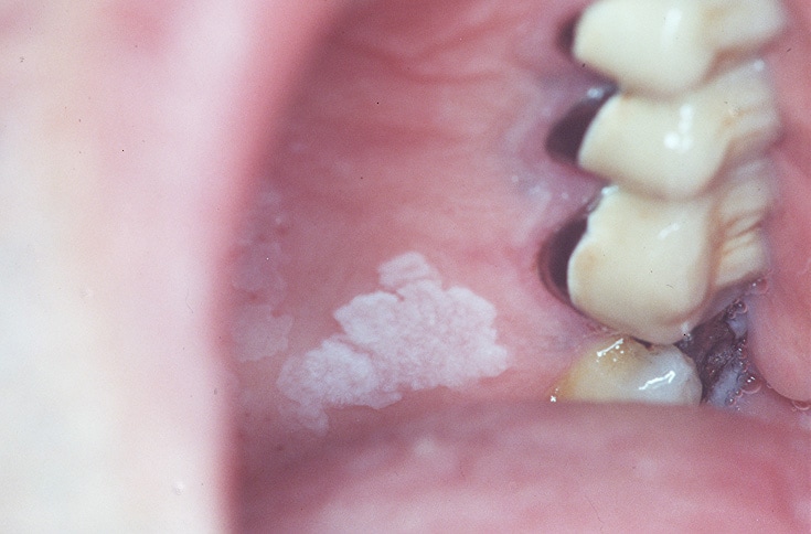
- Most leukoplakias are smooth, white plaques (homogeneous leukoplakias), as shown in the image below.
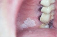
Homogeneous leukoplakia.
- Most leukoplakias occur on the lip, the buccal mucosae, or the gingivae.
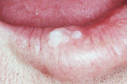
- Some leukoplakias are white and warty (verrucous leukoplakia), as shown in the image below.
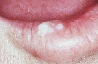
Verrucous or nodular leukoplakia.
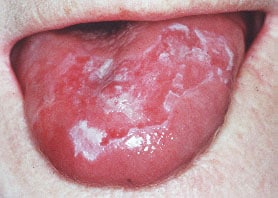
- Some leukoplakias are mixed white and red lesions (erythroleukoplakias or speckled leukoplakias), as shown in the image below.
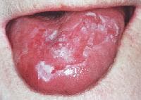
Erythroleukoplakia.
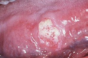
- Dysplastic lesions do not have any specific clinical appearance; however, where erythroplasia is present, dysplasia, carcinoma in situ, and frank carcinomas are more likely to be seen. The site of the lesion is relevant; leukoplakias on the floor of the mouth or on the ventrum of the tongue and the lip are sinister. The size of the lesion appears to be irrelevant. Even small dysplastic lesions may lead to multiple carcinomas and a fatal outcome. Note the image below.
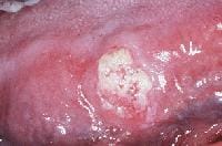
Carcinoma referred to as a leukoplakia.
Causes
No etiologic factor can be identified for most persistent oral leukoplakias (idiopathic leukoplakia). Known causes of leukoplakia include the following:
- Trauma (eg, chronic trauma from a sharp or broken tooth or from mastication may cause keratosis)
- Tobacco use: Chewing tobacco is probably worse than smoking.5
- Alcohol
- Use of betel, kat (Qat), and similar products6,7
- Infections (eg, candidosis, syphilis, Epstein-Barr virus infection): Epstein-Barr virus infection causes a separate and distinct non–premalignant lesion termed hairy leukoplakia.
- Chemicals (eg, sanguinaria)8,9
- Immune defects: Leukoplakias appear to be more common in transplant patients.
More on Leukoplakia, Oral |
 Overview: Leukoplakia, Oral Overview: Leukoplakia, Oral |
| Differential Diagnoses & Workup: Leukoplakia, Oral |
| Treatment & Medication: Leukoplakia, Oral |
| Follow-up: Leukoplakia, Oral |
| Multimedia: Leukoplakia, Oral |
| References |

Great post and thanks for posting it and sharing this..
Trả lờiXóawhite patch surgery in lucknow
I was diagnose with genital warts since 2012 i have be taking lot treatment and all i got is outbreak. in 2015 I gave up the treatment because I can't continues wasting time and money on treatment at the end it will not cure me. about 6 weeks ago i did natural research online I had So many people talking good about natural remedy, after the research i was recommended to Dr onokun, And I wrote to him through his email and told him my problem after some conversations with him he gave me natural treatment after 1 week Dr onokun treated me i got cured permanently. and i went to see my doc he confirmed that the diseases has gone out from my body. every patients should know there is 100% natural hpv cure. contact Dr onokun his email address: dronokunherbalcure@gmail.com
Trả lờiXóa