History of the Procedure
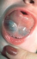
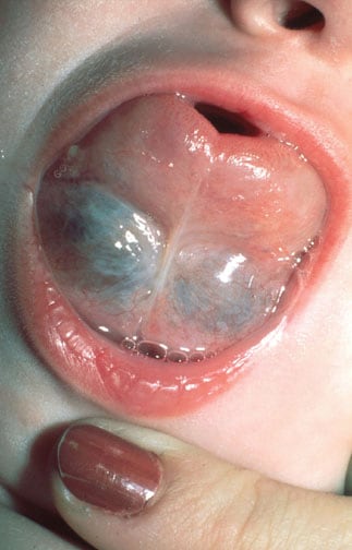
The term ranula is derived from the Latin word rana, meaning frog, and describes a blue translucent swelling in the floor of the mouth reminiscent of the underbelly of a frog. Hippocrates described ranulas and thought that they were secondary to inflammation. Paré thought that ranulas may represent descent of brain or pituitary matter.
An image depicting a ranula can be seen below.
Ranula. Image courtesy of Sylvan Stool, MD.
Frequency
Ranulas rarely occur. In one study of 1303 salivary gland cysts, only 42 were ranulas. The reported male-to-female ratio is 1:1.3, without significant side preference. Presentation is most frequently in the second and third decades of life, with an age range of 3-61 years.
Plunging ranulas occur less commonly than ranulas. Only slightly more than 100 well-documented cases of plunging ranulas have been reported in the English literature.
Etiology
Ranulas
Congenital ranulas can arise secondary to an imperforate salivary duct or ostial adhesion. These are very rare and have been known to spontaneously resolve.
Posttraumatic ranulas arise from trauma to the sublingual gland, leading to mucus extravasation and formation of a pseudocyst. The more appropriate term for this may be mucus escape reaction (MER).
Plunging ranulas
Other terms include deep, diving, cervical, or deep plunging ranula and oral ranula with cervical extension.
Plunging ranulas generally appear in conjunction with an oral ranula. Rarely, they can arise independently of the oral component. Patients present first with an oral swelling in up to 45% of cases, with associated oral swelling in 34%, and without any oral involvement in 21% of cases.
A 2010 review by Morton et al found evidence that suggested a genetic basis for plunging ranulas.1
Pathophysiology
Ranulas
Ranulas are formed from 1 of 2 processes:
- Partial obstruction of a sublingual duct can lead to formation of an epithelial-lined retention cyst. This is unusual, occurring in less than 10% of all ranulas.
- Trauma can lead to formation of ranulas. Experimentally, partial severance or ligation of the sublingual duct leads to ranula formation, whereas ligation of the submandibular duct does not. The ligation of the parotid duct ultimately leads to atrophy. The difference lies in the fact that the sublingual gland secretes continuously in the interdigestive period, whereas the other two major salivary glands only secrete in response to stimuli, such as eating. Therefore, with trauma, if a duct is obstructed, secretory backpressure builds and acini rupture, leading to mucus extravasation. Alternately, trauma causes direct damage to the duct or acini, leading to mucus extravasation. A pseudocyst then forms.
Plunging ranulas
Plunging ranulas arise in the neck by 3 mechanisms:
- The sublingual gland may project through the mylohyoid, or an ectopic sublingual gland may exist on the cervical side of the mylohyoid. This explains most plunging ranulas that exist without an oral component.
- The cyst may penetrate through the mylohyoid. Up to 27-45% of mylohyoid muscles in cadavers are found to be dehiscent, usually in the anterior two thirds of the muscle. These sites of dehiscence provide a route of egress for the cyst. In some instances, surgical trauma from initial ranula operations may scar or fibrose the superior surface of a ranula. When the ranula recurs, the path of least resistance is through a dehiscent mylohyoid, and a plunging ranula forms when only a simple ranula was present initially. Up to 44% of all plunging ranulas are iatrogenically induced in this manner.
- A duct from the sublingual gland may join the submandibular gland or its duct, allowing ranulas to form in continuity with the submandibular gland. Therefore, the ranula accesses the neck from behind the mylohyoid muscle.
Presentation
Ranulas
A ranula is most commonly observed as a bluish cyst located below the tongue as seen in the images below. It may fill the mouth and raise the tongue. Typically, these are painless masses that do not change in size in response to chewing, eating, or swallowing. Occasionally, pain may be involved.
Ranula. Image courtesy of Sylvan Stool, MD.

Ranula. Image courtesy of Sylvan Stool, MD.
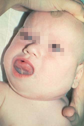
Plunging ranulas
Plunging ranulas can manifest as neck swelling in conjunction with, or independent of, a floor-of-mouth cyst. Occasionally, squeezing the mass causes swelling in the floor-of-mouth cyst. Most reported plunging ranulas are 4-10 cm in size and are usually found in the submandibular space. They have been reported to extend into the submental region, the contralateral neck, the nasopharynx up to the skull base, the retropharynx, and even into the upper mediastinum.
Indications
See Surgical therapy.
Relevant Anatomy
The sublingual gland lies against the sublingual depression of the mandible and directly on the mylohyoid. The submandibular duct (Wharton duct) and the lingual nerve lie medial to the gland. The genioglossus muscle is medial to these structures. No posterior fascial limits to the sublingual space exist, which allows lesions to exit the sublingual space and enter into the submandibular or parapharyngeal space.
Contraindications
Although some have advocated surgical management of congenital ranulas, recent literature supports observation in asymptomatic patients. Many congenital ranulas resolve on their own and do not require surgical intervention.
More on Ranulas and Plunging Ranulas |
 Overview: Ranulas and Plunging Ranulas Overview: Ranulas and Plunging Ranulas |
| Workup: Ranulas and Plunging Ranulas |
| Treatment: Ranulas and Plunging Ranulas |
| Follow-up: Ranulas and Plunging Ranulas |
| Multimedia: Ranulas and Plunging Ranulas |
| References |

I was diagnose with genital warts since 2012 i have be taking lot treatment and all i got is outbreak. in 2015 I gave up the treatment because I can't continues wasting time and money on treatment at the end it will not cure me. about 6 weeks ago i did natural research online I had So many people talking good about natural remedy, after the research i was recommended to Dr onokun, And I wrote to him through his email and told him my problem after some conversations with him he gave me natural treatment after 1 week Dr onokun treated me i got cured permanently. and i went to see my doc he confirmed that the diseases has gone out from my body. every patients should know there is 100% natural hpv cure. contact Dr onokun his email address: dronokunherbalcure@gmail.com
Trả lờiXóa