Introduction
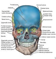
The facial skeleton serves to protect the brain; house and protect the sense organs of smell, sight, and taste; and provide a frame on which the soft tissues of the face can act to facilitate eating, facial expression, breathing, and speech. The primary bones of the face are the mandible, maxilla, frontal bone, nasal bones, and zygoma. Facial bone anatomy is complex yet elegant in its suitability to serve a multitude of functions.
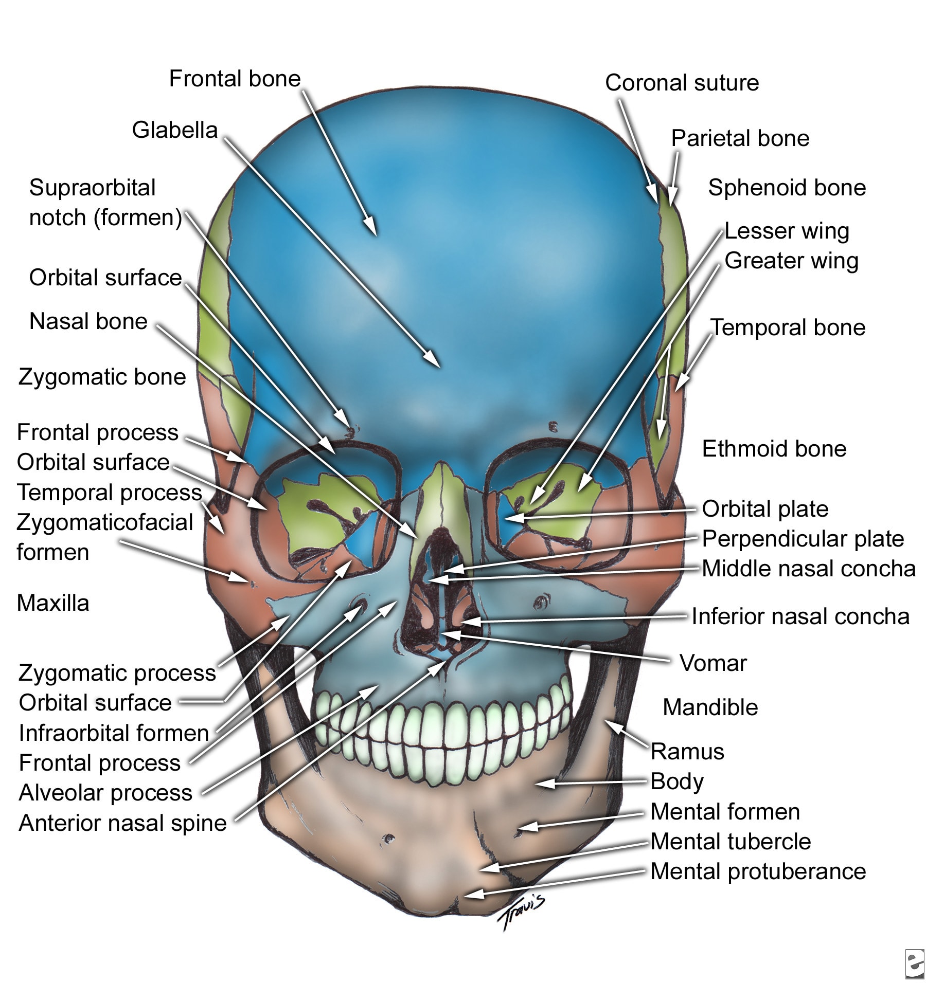
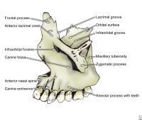
Skull, anterior view.
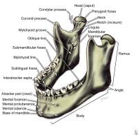
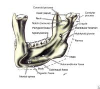
Mandible
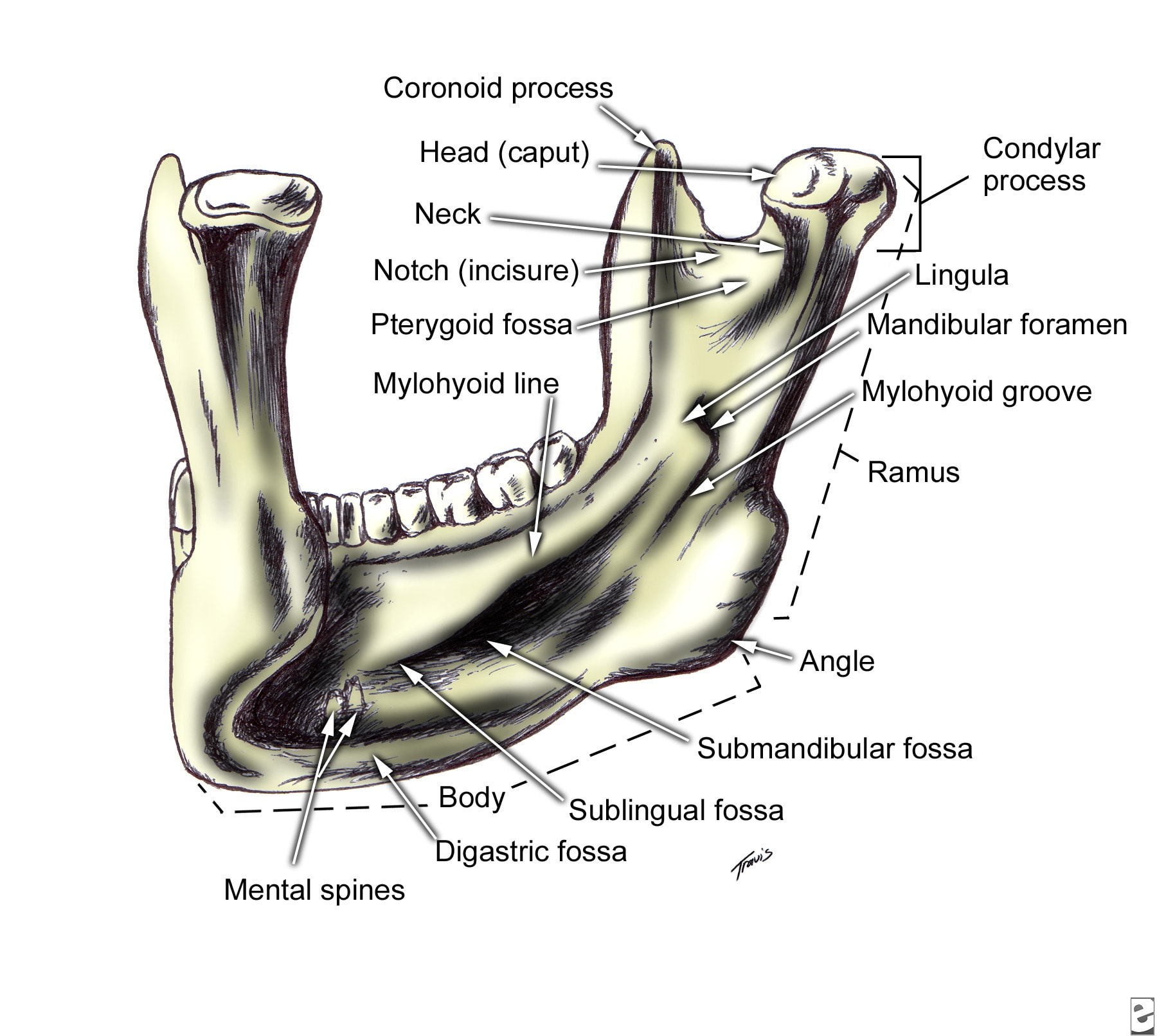
The mandible is a U-shaped bone. It is the only mobile bone of the facial skeleton, and, since it houses the lower teeth, its motion is essential for mastication. It is formed by intramembranous ossification. The mandible is composed of 2 hemimandibles joined at the midline by a vertical symphysis. The hemimandibles fuse to form a single bone by age 2 years. Each hemimandible is composed of a horizontal body with a posterior vertical extension termed the ramus.
Mandible, anterolateral superior view.

Mandible, left posterior view.
Body - Lateral surface
On the anterior inferior midline region of the hemimandible body is a triangular thickening of bone termed the mental protuberance. The thickened inferior rim of the mental protuberance extends laterally from the midline and forms 2 rounded protrusions termed the mental tubercles. Located lateral to the midline on the external surface are the mental foramina that transmit the mental nerves and vessels. They usually are located below the apex of the second bicuspid and have 6-10 mm of variation in the anteroposterior dimension. The rim of bone lateral to the mental tubercles extends posteriorly and ascends obliquely as the oblique line to join the anterior edge of the coronoid process. The inferior rim of the posterior body thickens and flares laterally where it attaches to the masseter muscle.
Body - Medial surface
Just lateral to the symphysis on the inner surface of the mandible are 2 paired protuberances termed the superior and inferior mental spines. The genioglossus muscle attaches to the superior mental spines, and the geniohyoid muscle attaches to the inferior mental spines. Just lateral to the inferior mental spines on the inferior border of the mandible are 2 concavities called the digastric fossae, where the anterior digastric muscles attach. Extending obliquely in a posterosuperior direction from the midline is a ridge of bone called the mylohyoid line, which serves as the attachment site for the mylohyoid muscle. Above and below the mylohyoid line on the inner mandibular body are 2 shallow convexities against which the sublingual and submandibular glands abut, respectively. Medial to the ascending edge of the anterior ramus is the retromolar trigone, located immediately behind the third molar.
Rami - Lateral surface
The ramus extends vertically in a posterosuperior direction posterior to the body on each hemimandible. The mandibular angle is formed by the intersection of the inferior rim of the body and the posterior rim of the ascending ramus. The superior ramus bifurcates into an anterior coronoid process and a posterior condylar process. The concavity between the 2 processes is called the mandibular notch. The coronoid is thin and triangular. With the teeth in occlusion, its superior extent is medial to the zygomatic arch. The coronoid is the site of attachment of the temporalis muscle. Inferiorly, the condylar process has a narrow neck that widens to a globular head that articulates with the glenoid fossa of the temporal bone.
Rami - Medial surface
On the medial surface of the ramus, just below the mandibular notch, is an aperture termed the mandibular foramen; the inferior alveolar nerve and blood vessels run through this aperture. Just medial to the mandibular foramen is the lingula, a triangular bony protuberance with its apex pointing posterosuperiorly toward the condylar head. Extending anteriorly and inferiorly from the mandibular notch toward the inferior rim of the body is the mylohyoid groove, through which the mylohyoid nerve runs.
The mandible has a large medullary core with a cortical rim 2-4 mm thick. The inferior alveolar canal begins at the mandibular foramen and courses inferiorly, anteriorly, and toward the lingual surface in the ramus. In adults, the canal comes in close proximity to the roots of the third molar. In the mandibular body, the canal courses along the inferior border close to the lingual surface. Anteriorly, the canal runs typically inferior to the level of the mental foramen, to which it ascends at its terminal end. The mandible houses the lower dentition, which in adults consists of 2 central and 2 lateral incisors, 2 canines, 2 first and 2 second premolars, and 3 sets of molars. Interdental septi run between the buccal and lingual cortices of the mandible, and interradicular septi run between the mesial and distal roots of the molars.
Maxilla
The maxilla has several roles. It houses the teeth, forms the roof of the oral cavity, forms the floor of and contributes to the lateral wall and roof of the nasal cavity, houses the maxillary sinus, and contributes to the inferior rim and floor of the orbit. Two maxillary bones are joined in the midline to form the middle third of the face.
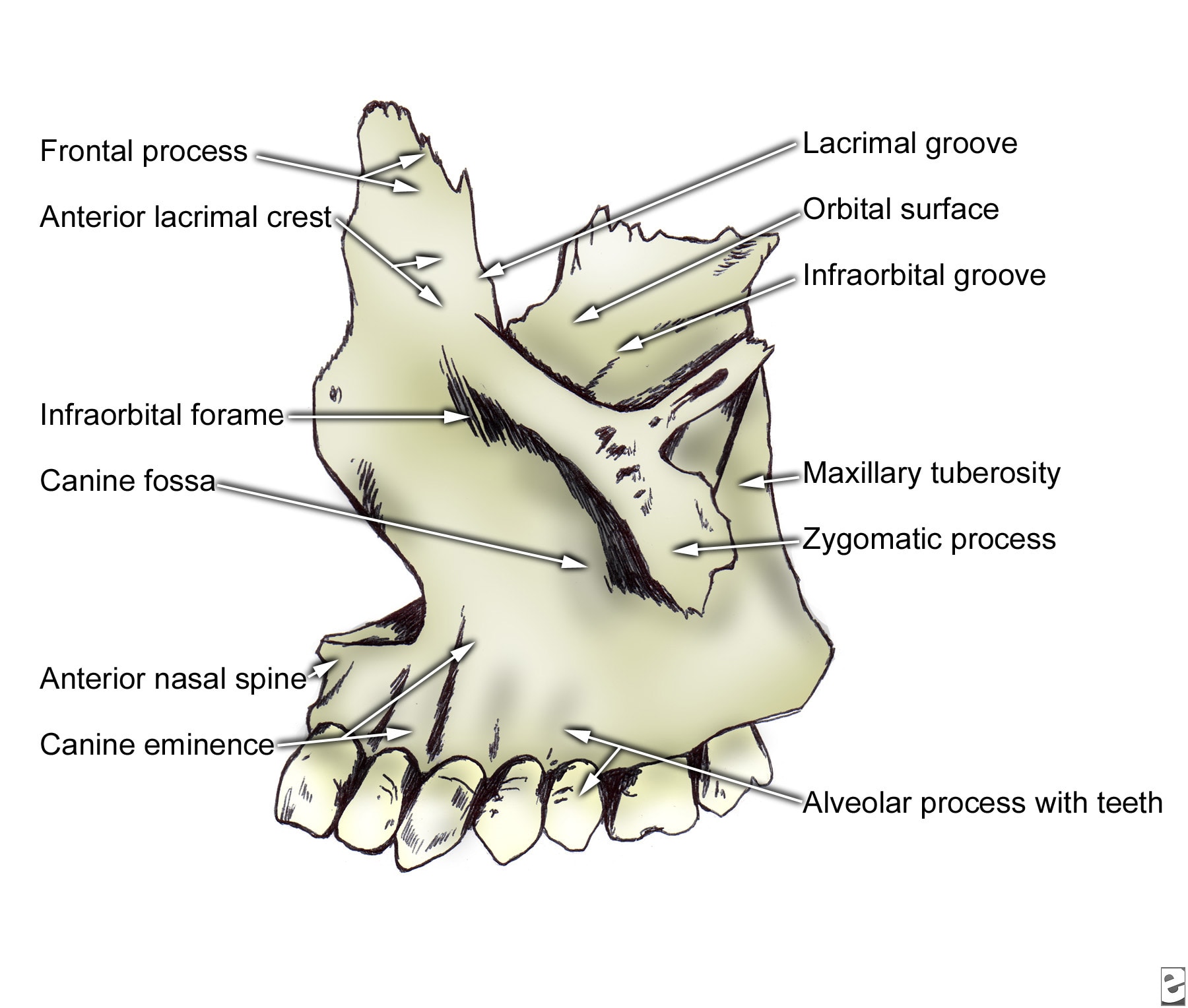
Left maxilla.
Anterior surface
In the midline of the anterior surface of the maxilla is found a prominence called the anterior nasal spine with a lateral concave rim, called the nasal notch, that forms the floor of the piriform aperture. Inferiorly, the alveolar process of the maxilla houses the teeth, including central incisors, lateral incisors, canines, 2 premolars, and 3 molars in adults. The tooth roots form vertical wavelike eminences in the anterior face of the maxilla; the canine root is the most prominent. The canine root forms a vertical ridge, termed the canine eminence, in the anterior face of the maxilla. The shallow fossae medial and lateral to the canine eminence are called the incisive fossa and the canine fossa, respectively.
Infraorbital rim and frontal process
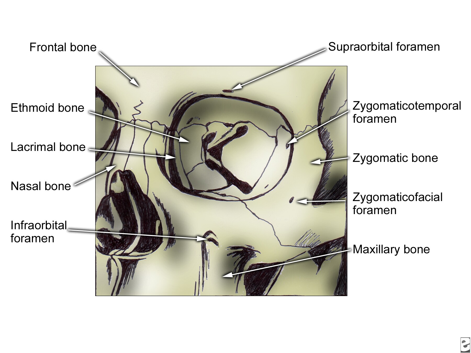
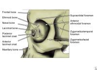
Superiorly, the maxillary bone is thickened in an inferior concavity that forms the infraorbital rim. Approximately 5-7 mm inferior to the rim lies the infraorbital foramen, which transmits the infraorbital nerve and vessels. The infraorbital rim extends medially and upward to form the frontal process of the maxilla. The frontal process articulates superiorly with the frontal bone, medially with the nasal bone, and posteriorly with the lacrimal bone. It has a smooth orbital surface that forms the vertical anterior lacrimal crest. Immediately posterior to the anterior lacrimal crest is a groove that forms the nasolacrimal canal.
Lateral portion
The maxilla projects laterally to form the zygomatic process, which articulates with the zygoma to form the lateral portion of the inferior orbital rim. Viewed medially, the maxillary sinus is evident with its medially facing ostium. It articulates with the palatine bone posteriorly and with the ethmoid, lacrimal, and inferior concha bones medially. In front of the maxillary sinus is a vertical nasolacrimal groove that forms the nasolacrimal canal with the lacrimal bone posteriorly and terminates inferiorly under the attachment of the inferior concha.
Superior surface
The superior surface of the maxilla forms the medial floor of the orbit. Posteriorly, the free edge forms the anterior border of the inferior orbital fissure. Medially, the orbital surface articulates with the ethmoid bone and lacrimal bone. Behind the frontal process of the maxilla and its anterior lacrimal crest is the nasolacrimal groove. Laterally, the orbital surface articulates with the orbital surface of the zygoma. On its inferior surface, the maxilla has a horizontal palatine process that forms the bulk of the hard palate. The palatine processes of both maxillae articulate with each other in the midline and with the horizontal plate of the palatine bone posteriorly. Anteriorly in the midline articulation of both palatine processes is the incisive canal, which transmits the nasopalatine nerve and branches of the greater palatine vessels. From a medial view, the maxillary hiatus is evident, opening into the maxillary sinus that occupies the predominant portion of the body of the maxilla.
Zygoma
The zygoma forms the lateral portion of the inferior orbital rim, as well as the lateral rim and lateral wall of the orbit.
Zygoma, frontal view.
Zygoma, lateral view.

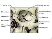
Additionally, it forms the anterior zygomatic arch, from which the masseter muscle is suspended. The masseter muscle acts to close the mandible for mastication and speech. On its lateral surface, the zygomatic bone has 3 processes. Inferiorly, a concave process projects medially to articulate with the zygomatic process of the maxilla, forming the lateral portion of the infraorbital rim. This concavity projects superiorly to form the frontal process that articulates with the frontal bone. Posteriorly, a temporal process articulates with the zygomatic process of the temporal bone to form the zygomatic arch. On the medial surface of the zygoma is a smooth orbital plate that forms the lateral floor and lateral wall of the orbit. It articulates posteriorly with the greater wing of the sphenoid bone.
Just posterior to the lateral rim and slightly inferior to the frontozygomatic suture is the marginal tubercle of Whitnall, to which the lateral palpebral ligament attaches. On the smooth medial orbital surface are foramina, which transmit the zygomaticofacial and zygomaticotemporal nerves to their respective apertures on the lateral surface. The zygomaticofacial foramen is located just lateral to the lateral orbital rim at the junction of the frontal and maxillary processes. The zygomaticotemporal foramen is located on the posterior concave surface of the lateral orbital rim.
Frontal Bone
Anterior surface
The frontal bone forms the anterior portion of the cranium, houses the frontal sinuses, and forms the roof of the ethmoid sinuses, nose, and orbit.
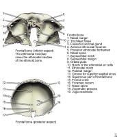
Frontal bone, inferior and posterior aspects.
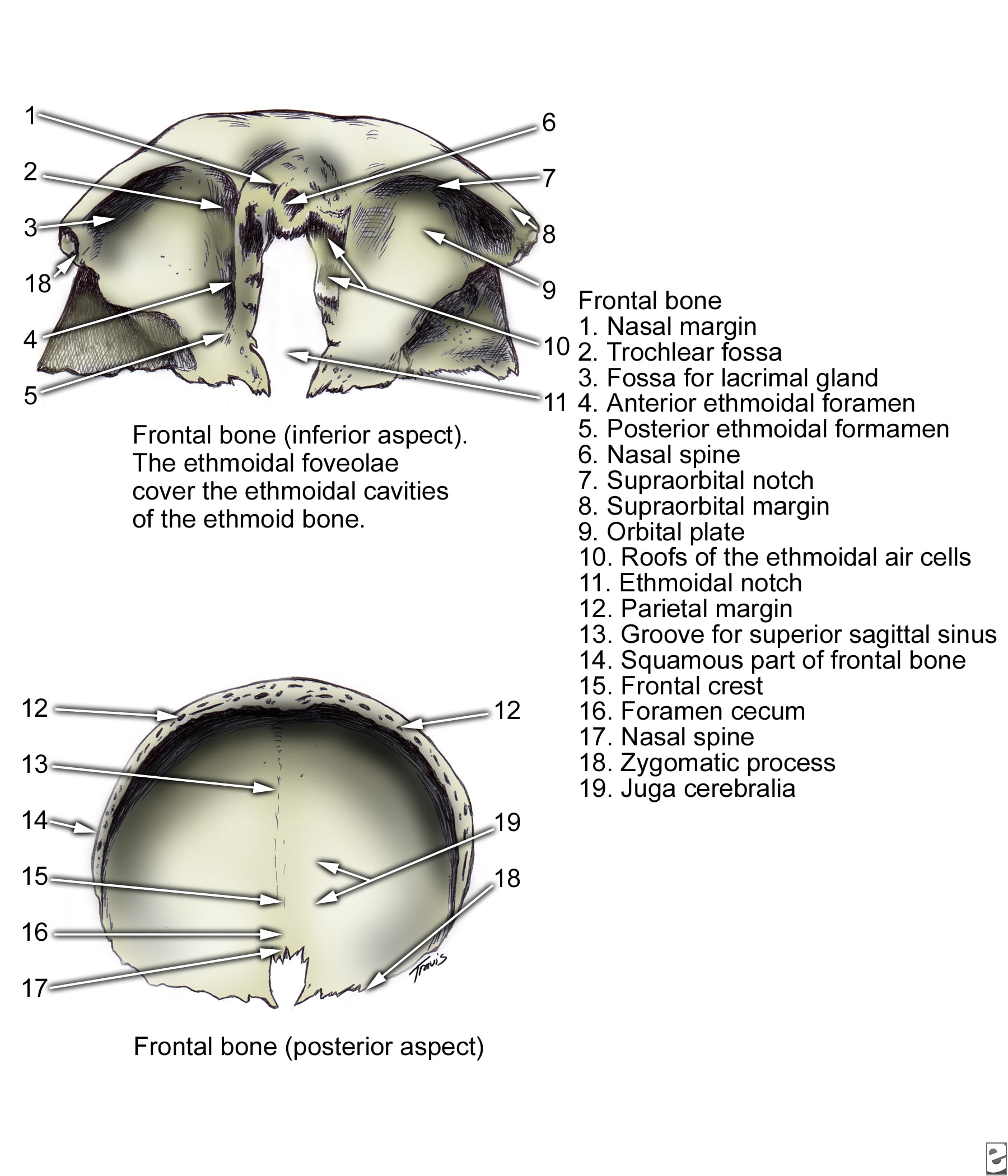
Anteriorly, the external surface is convex superiorly, and it articulates with the parietal bones posteriorly and the greater wing of the sphenoid posteroinferiorly. The anterior convex surface thickens inferiorly to form the supraorbital rims. Just above the supraorbital rims are thickened arches termed the superciliary arches. Their midline union forms a depression called the glabella. At the junction of the lateral third and the medial third of each supraorbital rim is either a notch or foramen through which the supraorbital vessels and nerves run. Bilateral notches are found in 49% of skulls, bilateral foramina in 26%, and 1 notch and 1 foramen in 24%.
Inferolaterally, the supraorbital ridge forms the zygomatic process that articulates with the frontal process of the zygomatic bone. The inferior surface of the frontal bone forms the concave surface of the orbital roof and the anterior nasal roof.
Orbital surface
The orbital surface articulates posteriorly with the greater and lesser wings of the sphenoid bone; medially with the ethmoid, lacrimal, and maxillary bones; and laterally with the zygoma. On the anterolateral orbital surface is a poorly developed depression for the lacrimal gland. On the medial orbital surface is a small mound where the cartilaginous trochlea of the superior oblique muscle attaches. Anteriorly, between the orbital surfaces, the frontal bone articulates with the anterior portions of the nasal bones and frontal processes of the maxilla. A small vertical midline plate, termed the nasal spine of the frontal bone, contributes to the nasal septum.
Internal surface
The internal surface of the frontal bone is concave anteriorly with grooves laterally for the middle meningeal vessels. The floor of the internal surface forms the floor of the anterior cranial fossa. It has a midline dehiscence termed the ethmoid notch that articulates with the ethmoid bone. Anterior to the ethmoid notch is an upward sloping ridge termed the frontal crest, which forms a groove for the superior sagittal sinus. At the anterior articulation between the frontal crest and the cribriform plate of the ethmoid bone is a small foramen called the foramen caecum, which usually transmits an emissary vein from the roof of the nasal cavity to the superior sagittal sinus.
Frontal sinus
The frontal sinus begins to form after age 3.5 years. It is located in the anterior midline between the supraorbital rims. In adults, the average dimensions reach 2.4 mm high, 2.9 cm to either side from the midline, and 2 cm in anteroposterior dimension. The anterior wall of the frontal bone splits to enclose the sinus, but both anterior and posterior walls retain their diploë. The frontal sinus usually is divided sagittally by a frequently eccentric septum. Just lateral to the midline, the bone of the anterior wall of the sinus is 4 mm thick, and the bone of the posterior wall is 1.9 mm thick.
Nasal Bones
The paired nasal bones form the anterosuperior bony roof of the nasal cavity. They are approximately quadrangular. They articulate with the nasal process of the frontal bone superiorly, the frontal process of the maxillary bone laterally, and with one another medially. Their inferior border is free and forms the superior margin of the piriform aperture. The external surface is convex except for the superior-most portion, where a concavity forms as the margin turns superiorly to articulate with the frontal bone. On the internal surface is a vertical groove for the external nasal artery.
Multimedia
 | Media file 1: Skull, anterior view. |
 | Media file 2: Mandible, anterolateral superior view. |
 | Media file 3: Mandible, left posterior view. |
 | Media file 4: Left maxilla. |
 | Media file 5: Zygoma, frontal view. |
 | Media file 6: Zygoma, lateral view. |
 | Media file 7: Frontal bone, inferior and posterior aspects. |
Keywords
facial bone anatomy, facial anatomy, bones of the face, mandible, maxilla, frontal bone, nasal bones, zygoma, facial skeleton, facial bones

I was diagnose with genital warts since 2012 i have be taking lot treatment and all i got is outbreak. in 2015 I gave up the treatment because I can't continues wasting time and money on treatment at the end it will not cure me. about 6 weeks ago i did natural research online I had So many people talking good about natural remedy, after the research i was recommended to Dr onokun, And I wrote to him through his email and told him my problem after some conversations with him he gave me natural treatment after 1 week Dr onokun treated me i got cured permanently. and i went to see my doc he confirmed that the diseases has gone out from my body. every patients should know there is 100% natural hpv cure. contact Dr onokun his email address: dronokunherbalcure@gmail.com
Trả lờiXóa